OCULUS
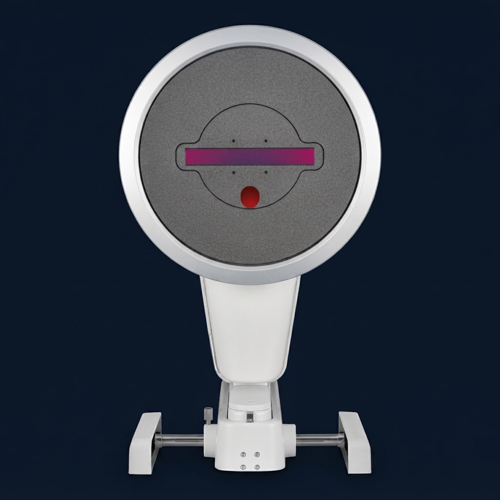
OCULUS Pentacam® Cornea OCT
Seeing Beyond Tomography
The Gold Standard. Now with Corneal Microstructure.
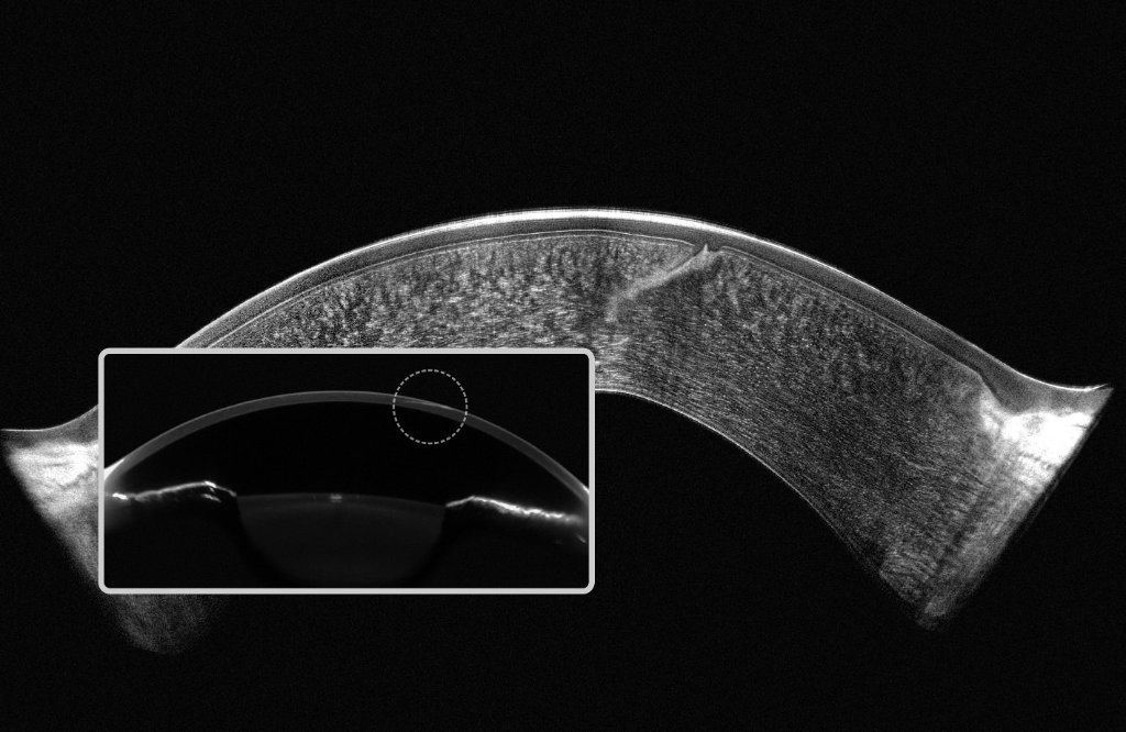
The OCULUS Pentacam® Cornea OCT combines the world's most trusted anterior segment tomography platform with ultra-high resolution 1.9 µm corneal OCT in a single, non-contact examination. Acquiring Scheimpflug tomography and OCT images simultaneously, it provides both comprehensive anterior segment measurements and microscopic visualisation of the cornea in just one scan.
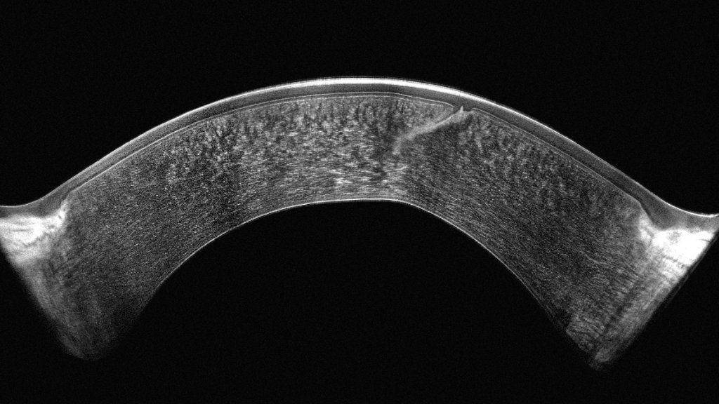
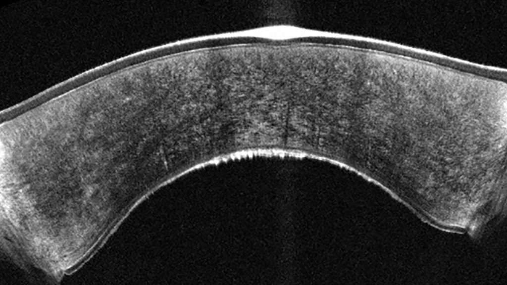
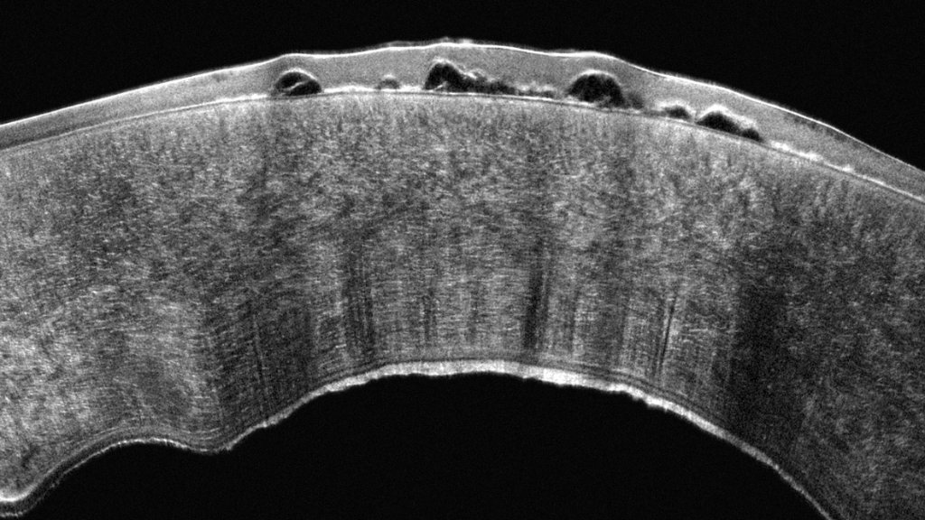
Designed for corneal, refractive and cataract specialists, the Pentacam Cornea OCT extends the proven clinical value of the Pentacam by allowing clinicians to assess not only corneal shape and biomechanics, but also the integrity of individual corneal layers including the tear film, epithelium, Bowman's layer and anterior stroma.
Whether screening refractive surgery candidates, evaluating keratoconus, planning premium cataract procedures or monitoring ocular surface disease, the Pentacam Cornea OCT provides a level of structural detail previously unavailable from tomography alone—supporting earlier diagnosis, greater surgical confidence and improved patient communication.
Combining Two Technologies in One Single Device
The Pentacam® Cornea OCT unites proven Scheimpflug tomography with ultra-high-resolution corneal OCT — capturing both datasets in a single, non-contact measurement for the most complete anterior segment assessment available.
Scheimpflug Tomography
The proven gold-standard rotating Scheimpflug camera captures the complete anterior segment — corneal front and back surfaces, anterior chamber depth, volume and angle, and lens — delivering comprehensive tomographic data in under 2 seconds.
- Complete anterior segment analysis
- True elevation data front & back
- Keratoconus detection & staging
- IOL power calculation support
Corneal OCT
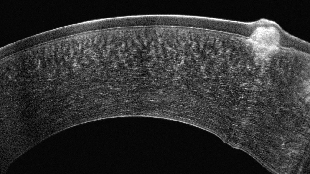
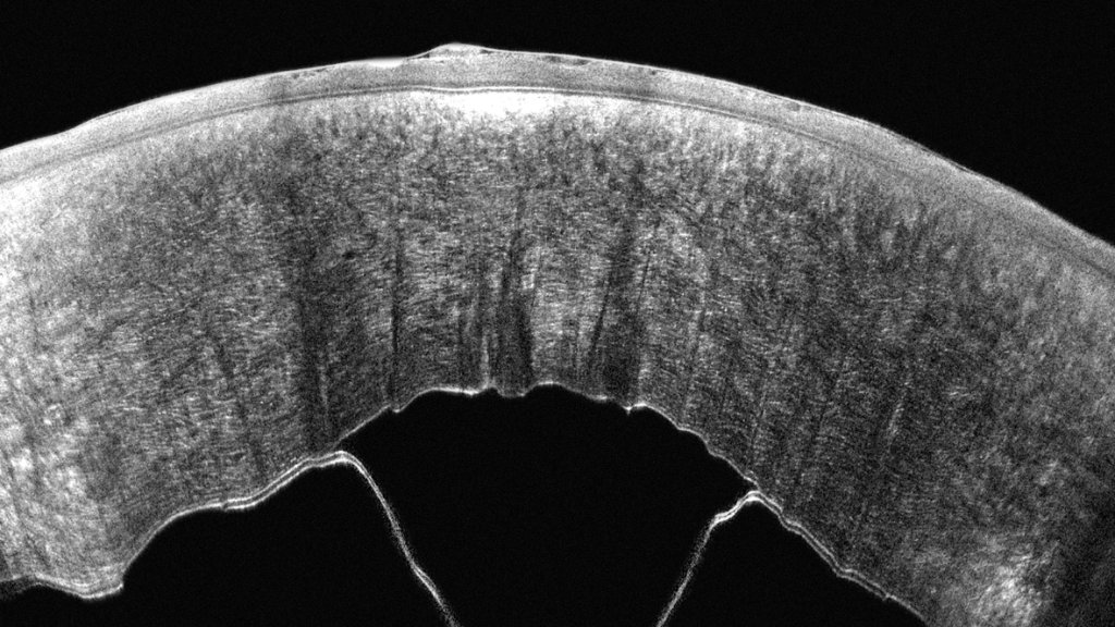
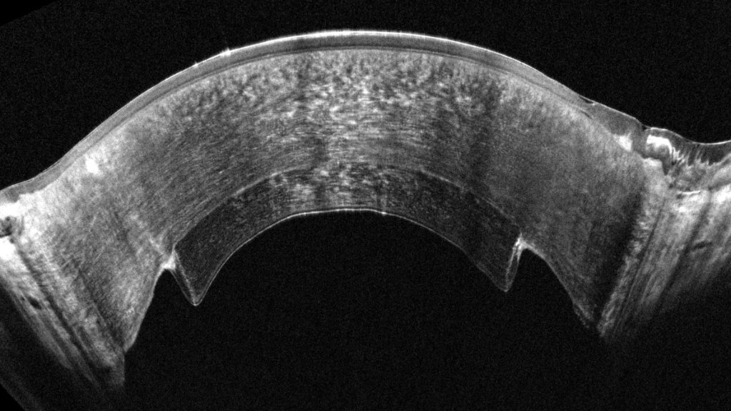
Ultra-high-resolution 1.9 µm OCT imaging visualises the cornea's microstructure — from the tear film through to deeper stromal layers — revealing structural detail invisible to tomography alone.
- 1.9 µm axial resolution
- Corneal layer segmentation
- Tear film thickness mapping
- Microstructural visualisation
One Measurement. Complete Confidence.
By acquiring both datasets simultaneously, the Pentacam Cornea OCT eliminates the need for multiple devices or patient repositioning — delivering comprehensive tomography and microscopic corneal detail in a single, time-saving examination.
See Pentacam Cornea OCT image quality